Featured Course
Point-of-Care Ultrasound for Primary Care
This course is designed for physicians, nurse practitioners, physician assistants and other allied health professionals interested in basic point-of-care ultrasound.
VIEW TRAINING COURSE
Case Study Details
| Patient Information | |
|---|---|
| Age | 54 |
| Gender | Male |
| Ethnicity | Hispanic |
| Symptoms (described) | No symptoms. Patient was referred for AAA assessment. |
| Additional Symptoms (observed) | |
| Health Status | CAD with recent percutaneous trans-coronary angioplasty. HTN, hyperlipidemia, ex-smoker (quit >40 years ago). Family hx, father died of ruptured AAA. |
| Evaluation | |
|---|---|
| Prior tests ordered | None |
| U/S ordered | Aorta duplex |
| Additional testing ordered | CTA 9/17/08, CT-guided Biopsy 10/2/08, Mesenteric Angiogram 10/23/08, Surgical Biopsy 11/12/08 |
| U/S performed by | Leah Jolly |
| Scanning Protocol | |
|---|---|
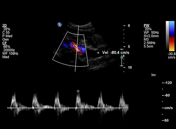
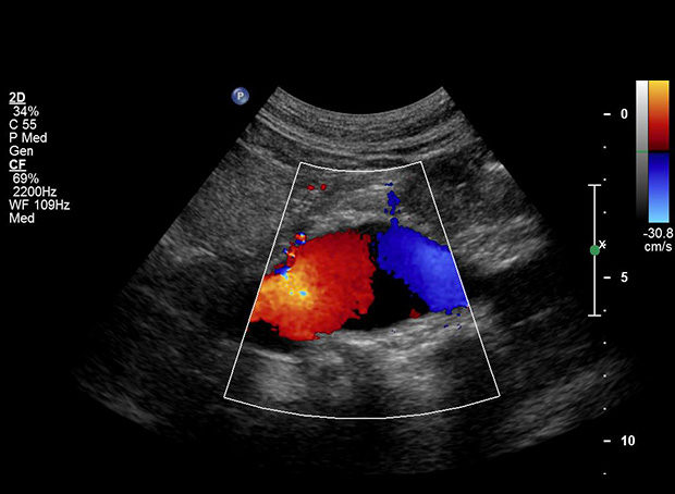
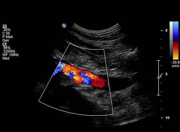
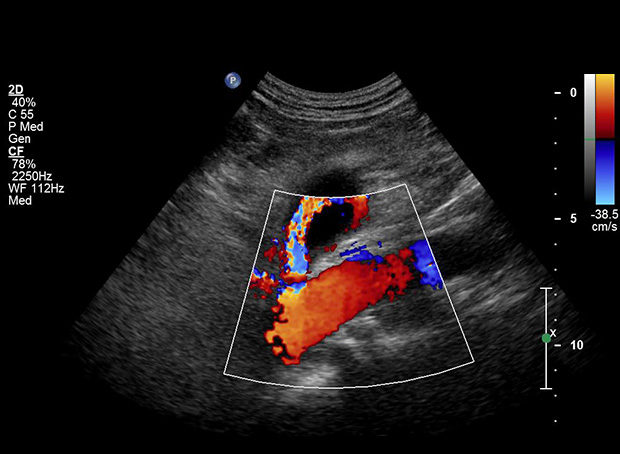
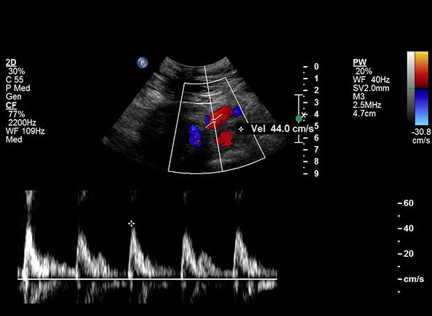
| Describe (where, when, how): | Abdominal aorta measured at proximal, mid, and distal segments in transverse and AP planes. Proximal common iliac arteries also measured. Doppler velocities obtained at all levels as well. Incidental note of a large round structure superior to the aorta. Further investigation performed with diameter and velocity measurements, as well as color flow images obtained. |
| What equipment was used? | Philips iU22 |
| What technique did you feel was most important for this exam? | Expanding the exam protocol when something out of the ordinary was noted. |
| Outcome | |
|---|---|
| Results | AAA 3.8 x 3.8 cm. Right CIA aneurysm 2.4 cm, left 1.8 cm. Incidental finding of superior mesenteric artery aneurysm measuring 3.5 x 3.0 cm with mural thrombus noted and lumen diameter 0.77 cm. |
| Note | CT revealed soft tissue density encompassing the SMA, possibly a thrombosed pseudoaneurysm versus neoplasm. CT Biopsy performed with inconclusive results. Mesenteric angiogram revealed normal caliber SMA with mild mass effect on the 1st and 2nd jejunal branches. Normal flow in all mesenteric vessels. Surgical biopsy performed, revealing low-grade B-Cell lymphoma |
| Diagnosis | Cancer – Non-Hodgkins Lymphoma |
Cancer is non-aggressive, so doctors and patient opted for surveillance rather than surgical treatment. He had one round of oral chemotherapy after a follow-up CT revealed mild progression. Mass shrunk with treatment. Now on maintenance Rituxan for 2 years.
Continues follow-up CT and PET scans as recommended by oncologist. AAA surveillance with ultrasound every 6 months.
Tell Us Your Story
We want to hear how you are using Ultrasound to improve patient care. Email Us Your Story